

 Image 1 of 3
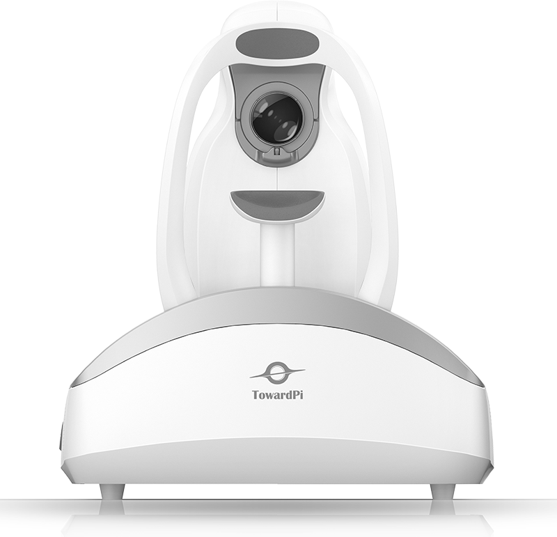 Image 2 of 3
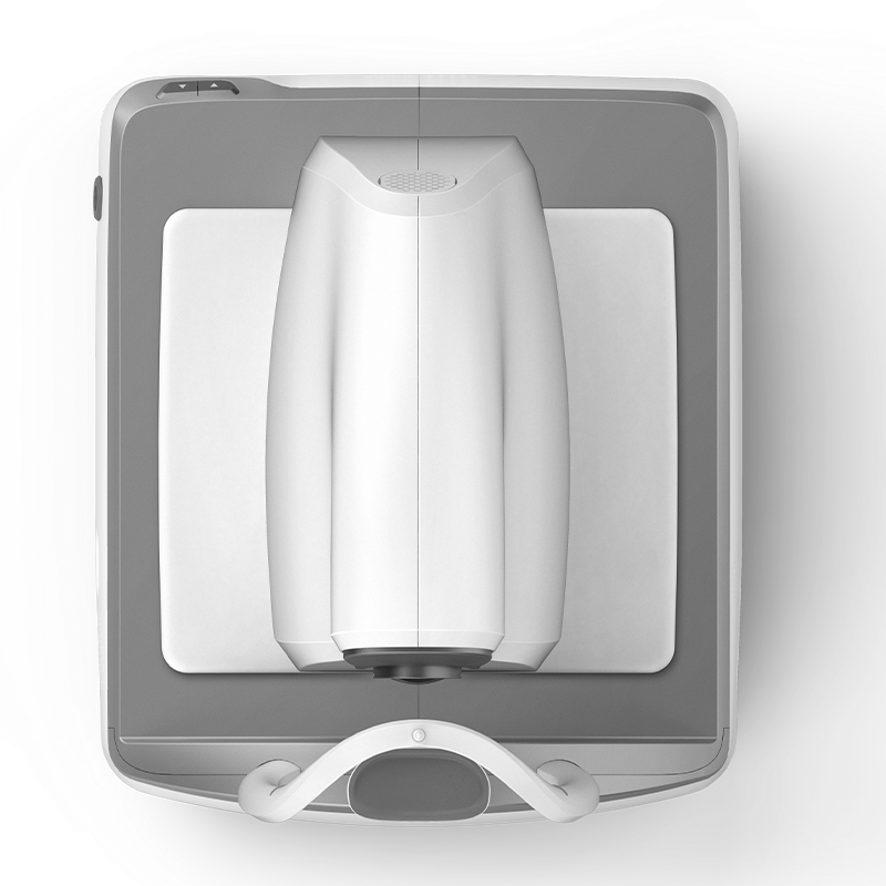 Image 3 of 3
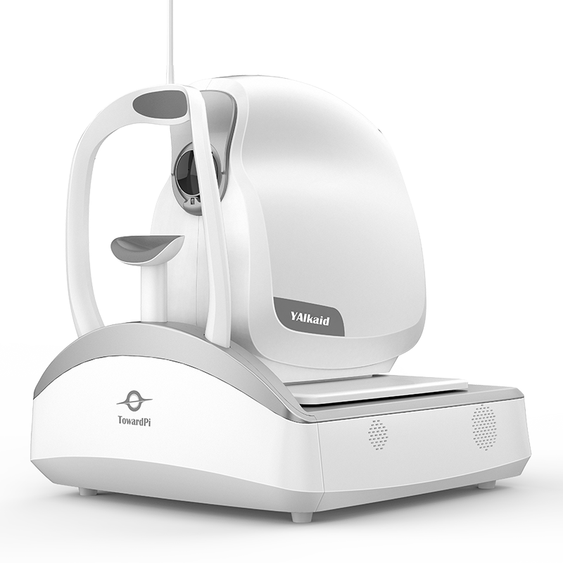
The TowardPi Yalkaid YG-250K is an ultra-widefield, full-range swept-source optical coherence tomography (SS-OCT) system designed for comprehensive imaging of both the anterior and posterior segments of the eye. With a scanning speed of 250,000 A-scans per second, it supports wide-area OCT and OCT angiography (OCTA) imaging, with scan widths of up to 30 mm for the posterior segment and 24 mm for the anterior segment, and a scan depth of up to 12 mm for both.
Operating at a central wavelength of 1060 nm, the YG-250K delivers an axial optical resolution of 3.8 μm. Its integrated scanning laser ophthalmoscopy (SLO) fundus imaging, 128 Hz eye tracking, and support for imaging through pupils as small as 2.0 mm help facilitate image acquisition across a range of examination conditions.
The system incorporates quantitative analysis tools for retinal and choroidal thickness and volume, blood flow, optic disc, glaucoma, and anterior segment parameters. Additional functions include choroidal OCTA algorithms, retinal curvature analysis, 3D structural imaging, and vascular analysis, supporting detailed assessment of ocular structures and blood flow.
