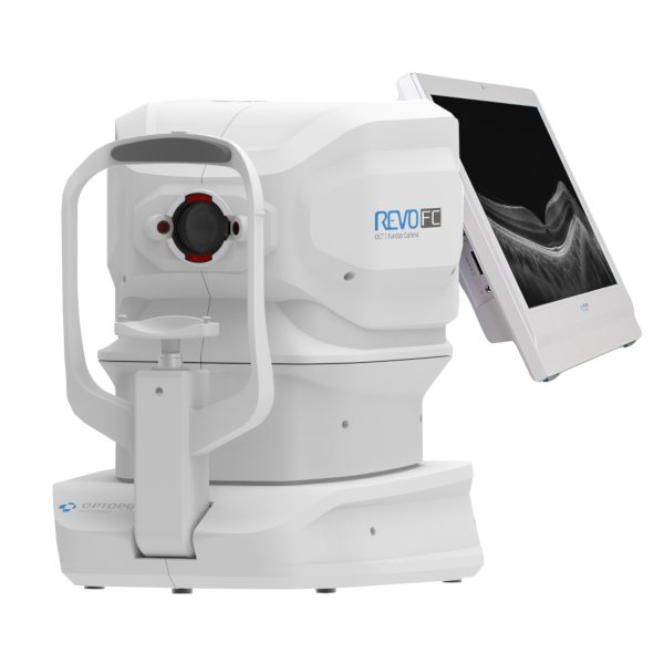
Optopol REVO FC Spectral-Domain OCT & Color Fundus Camera

SGD 0.00

The Optopol REVO FC is an all-in-one Spectral-Domain OCT and non-mydriatic colour fundus camera designed for comprehensive anterior and posterior segment imaging. Combining high-resolution OCT with automated retinal photography, the system provides a streamlined solution for retinal, glaucoma, and anterior segment assessment within a single diagnostic platform.

With a scanning speed of 80,000 A-scans per second, the REVO FC provides detailed OCT imaging with an axial resolution of up to 2.8 µm digitally. Full Range imaging extends the scan depth for challenging cases, including highly myopic eyes, while AccuTrack™ real-time hardware-based eye tracking helps compensate for blinks, fixation loss, and involuntary eye movements during acquisition.

The integrated 12.3 MP non-mydriatic colour fundus camera captures high-resolution retinal images with a 45° field of view and supports automated alignment, focusing, flash and image capture. The system also supports anterior segment photography, allowing documentation of corneal and anterior eye structures within the same workflow.

The Optopol REVO FC is an all-in-one Spectral-Domain OCT and non-mydriatic colour fundus camera designed for comprehensive anterior and posterior segment imaging. Combining high-resolution OCT with automated retinal photography, the system provides a streamlined solution for retinal, glaucoma, and anterior segment assessment within a single diagnostic platform.

With a scanning speed of 80,000 A-scans per second, the REVO FC provides detailed OCT imaging with an axial resolution of up to 2.8 µm digitally. Full Range imaging extends the scan depth for challenging cases, including highly myopic eyes, while AccuTrack™ real-time hardware-based eye tracking helps compensate for blinks, fixation loss, and involuntary eye movements during acquisition.

The integrated 12.3 MP non-mydriatic colour fundus camera captures high-resolution retinal images with a 45° field of view and supports automated alignment, focusing, flash and image capture. The system also supports anterior segment photography, allowing documentation of corneal and anterior eye structures within the same workflow.

Optional Software Modules


Enquire Now