

 Image 1 of 3
 Image 2 of 3
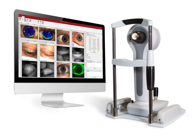 Image 3 of 3
-
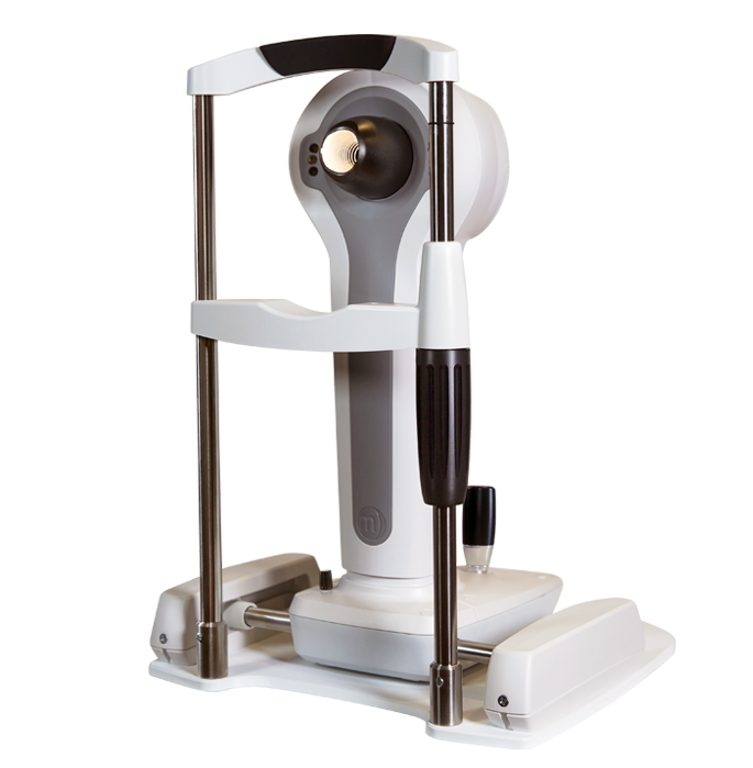
The Medmont Meridia delivers a large capture area with high-resolution imaging, providing more real corneal data in a single image for improved diagnostic precision and reduced estimation.
-
An ergonomic instrument design with integrated quick-access keys and software guidance tools supports efficient workflow, improved usability, and better space optimization within clinical environments.
-
The Professional model includes anterior imaging, fluorescein imaging, meibomian gland assessment, video capture, and detailed patient documentation tools to support comprehensive ocular surface evaluation.
-
Meaningful patient views, proven grading scales, and visual reports help improve patient understanding, consultation quality, treatment planning, and long-term patient engagement.

