

 Image 1 of 2
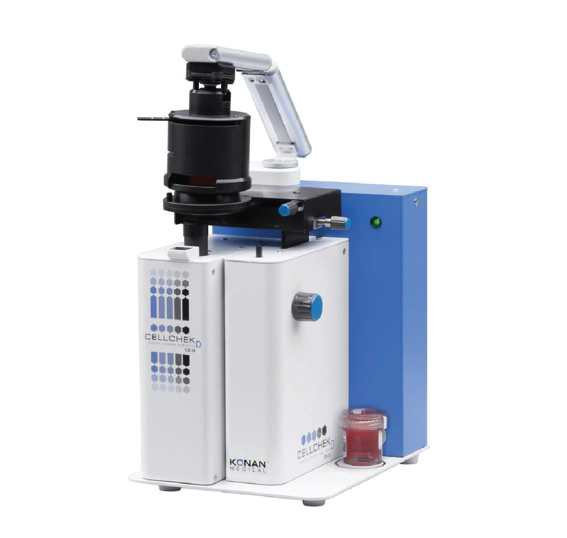 Image 2 of 2
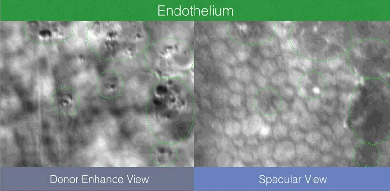
Donor Enhanced Imaging System
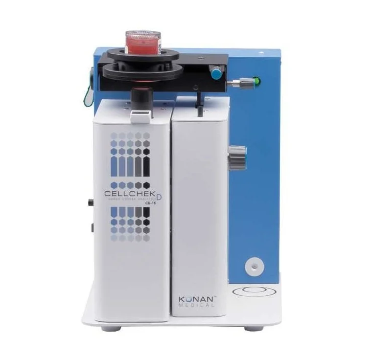
The CellChek D+ features an advanced “Donor Enhanced” imaging system that provides detailed visualization of the corneal endothelium, intra-stromal structures, and epithelium. This supports evaluation of features such as blood cells, epithelial anomalies, rough keratome cuts, dead cells, and potential fungal presence for improved donor tissue assessment.
Wide Viewing & Multi-Sample Analysis
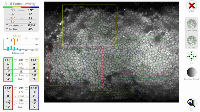
The system provides broad overview imaging together with large multi-sample analytical areas. Integrated finder and digital measurement tools support efficient documentation and corneal evaluation workflow.
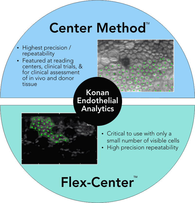
Patented Cellular Analysis Methods
Konan’s patented Center Method provides highly precise and repeatable endothelial cell analysis and is widely regarded as a gold-standard assessment method. The Flex-Center Method further supports analysis of advanced diseased corneas where only limited visible cells are present, enabling reliable evaluation across a wide spectrum of corneal conditions.
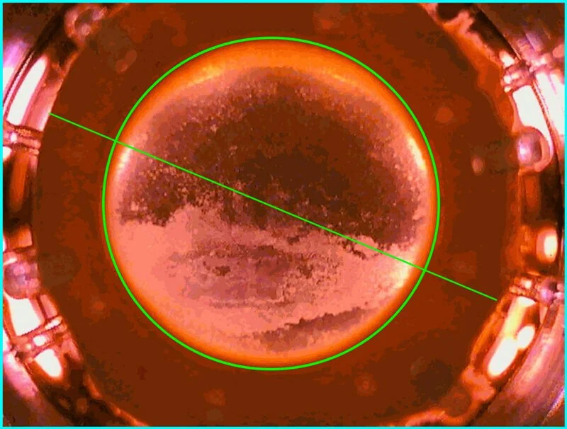
Full Graft Imaging & Documentation
CellChek D and D+ provide complete corneal imaging with digital measurement tools for identifying, measuring, and documenting corneal dimensions, scars, and defects.
