

 Image 1 of 2
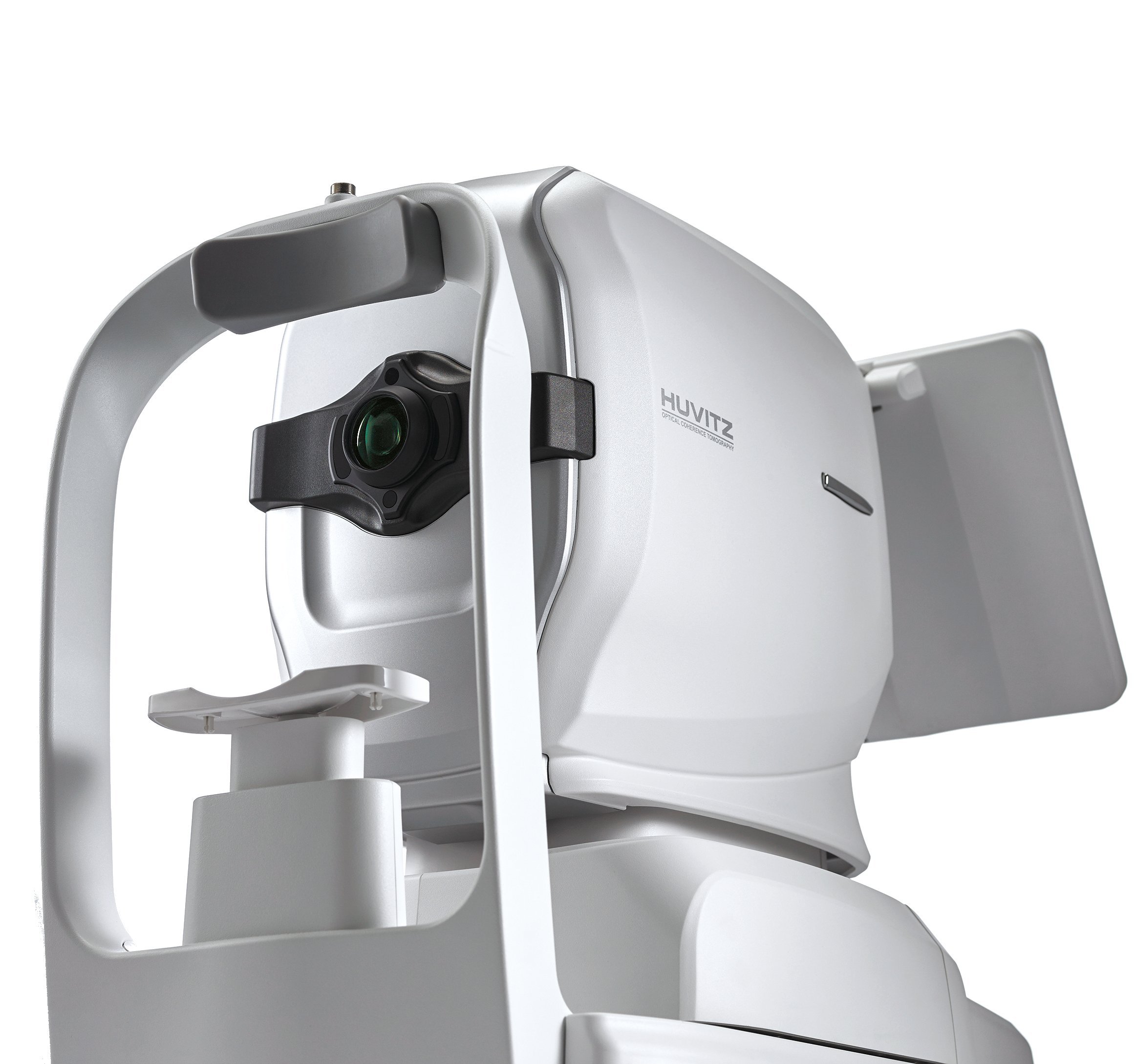 Image 2 of 2
High-Speed Imaging & High-Quality Visualization
The HOCT series delivers high-speed scanning and high-quality imaging through Huvitz’s advanced optical technology and image processing software. Detailed visualization of retinal structures, including macular thickness and layer separation, supports comprehensive ophthalmic assessment.
All-in-One Diagnostic System
By integrating OCT angiography, a full color fundus camera, and a built-in PC system, the HOCT enables multi-functional ophthalmic diagnosis within a single platform. Frontal view imaging, tomography, comparative analysis, and diagnostic evaluation can all be performed efficiently in one workflow.
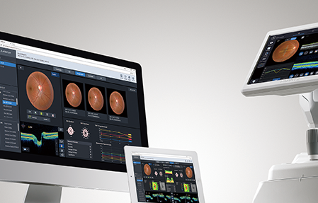
Web-Based Data Access
Patient examination data can be reviewed and analyzed remotely through standard web browsers including Chrome, Safari, and Internet Explorer without requiring dedicated software installation.
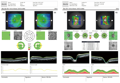
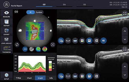
Comprehensive Reporting System
The system generates detailed and easy-to-read reports displaying pathological structures and clinically relevant analysis data. Reports can be viewed through web browsers and printed in multiple report formats directly from the analysis interface.
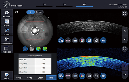
Anterior Segment Measurement
The anterior segment module supports measurement and analysis of corneal thickness, anterior chamber angle, and 3D anterior imaging. Both anterior and posterior segment assessments can be performed within the same platform for improved clinical efficiency. Optional 9 mm and 16 mm anterior lens sets are available.

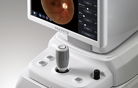
Full Color Fundus Imaging
The integrated full color fundus camera captures high-resolution retinal images with optimized contrast for enhanced clinical analysis and diagnosis. Features including low-intensity flash, fast image capture, quiet operation, small pupil mode, and automatic flicker detection improve image quality and patient comfort.
